Background: Leukoplakia of the vocal cords may represent a pre-cancerous lesion of the larynx. The management of cases of recurrent leukoplakia with pathologically proven dysplasia is still controversial. Objectives: To present a series of patients with recurrent vocal cord leukoplakia and to examine their malignant transformation rate in relation to the clinical characteristics, risk factors.. Table 2. Number and percentage distribution of ENT and pathology diagnoses of vocal fold nodules and polyps. In comparing the clinical and histological findings, there was agreement in 123 (93.18%) lesions from the 132 lesions analyzed, making up a total of 56 nodules (42.42%) and 67 polyps (50.76%).

Pathology Outlines Papilloma
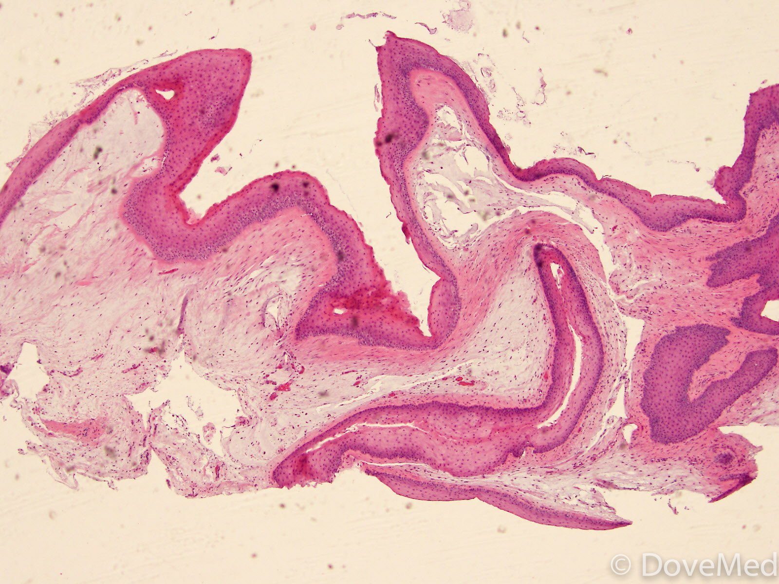
Pathology Outlines Vocal cord polyp
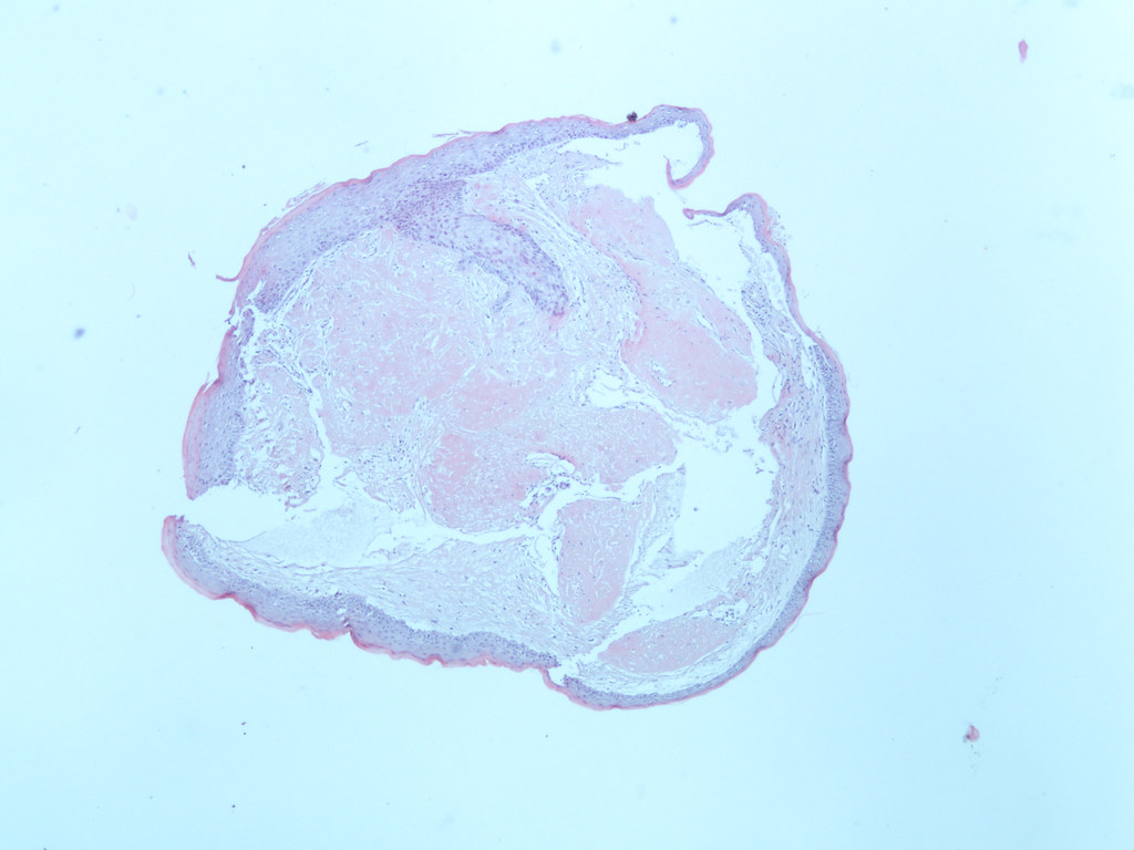
Pathology Outlines Vocal cord polyp
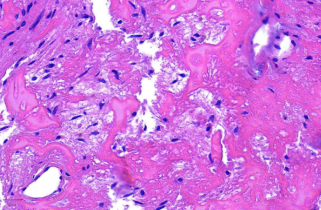
Pathology Outlines Vocal cord polyp
Vocal Cord Dysplasia (precancer) Symptoms, Causes, Treatment, and More Article Insider
Pathology Outlines Papilloma
Pathology Outlines Vocal cord polyp
vocal cord polyp vocal cord, larynx, hypopharynx histology
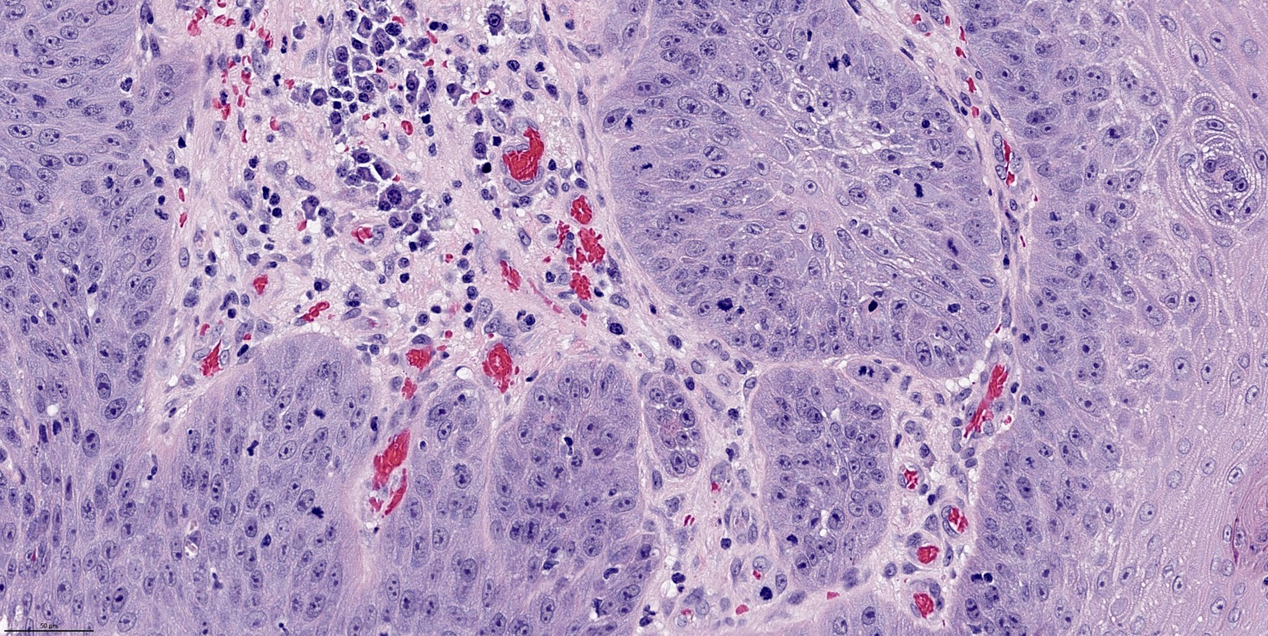
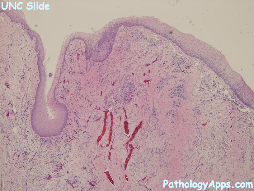
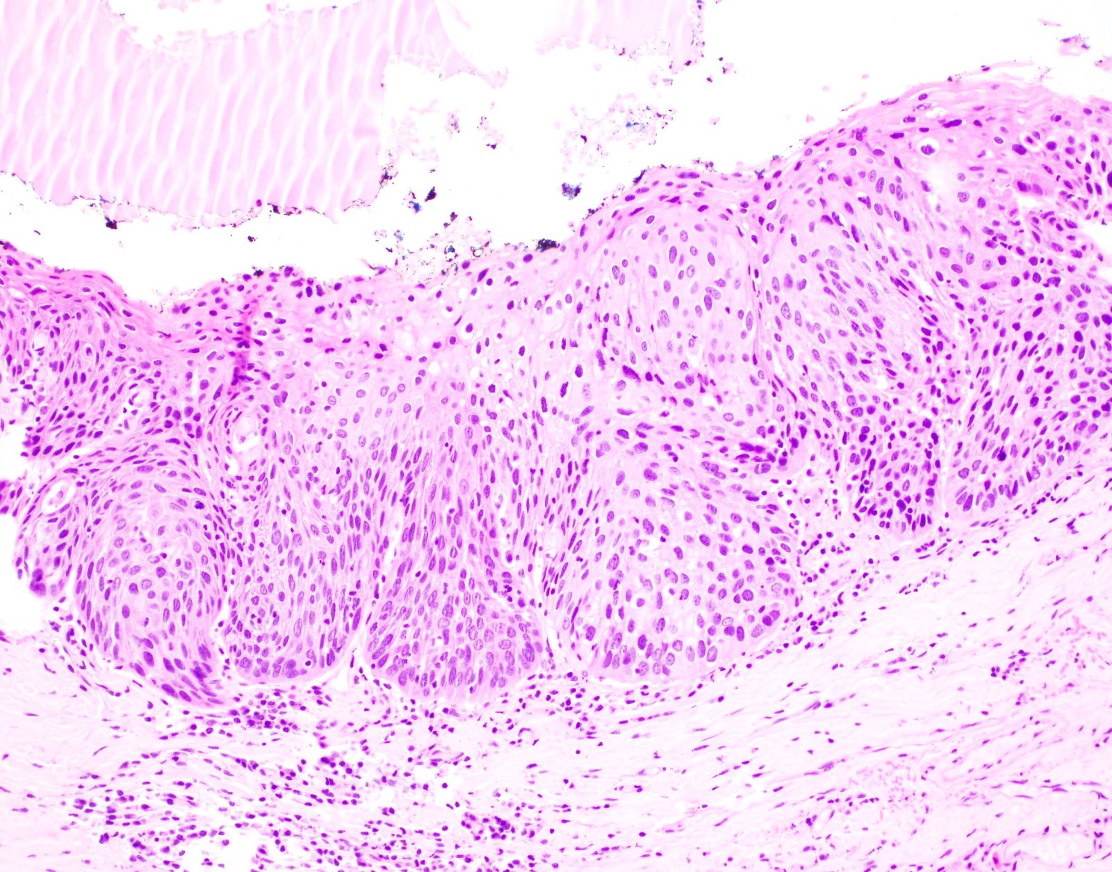
Severe squamous dysplasia or Carcinoma in situ causing laryngeal leukoplakia Iowa Head and
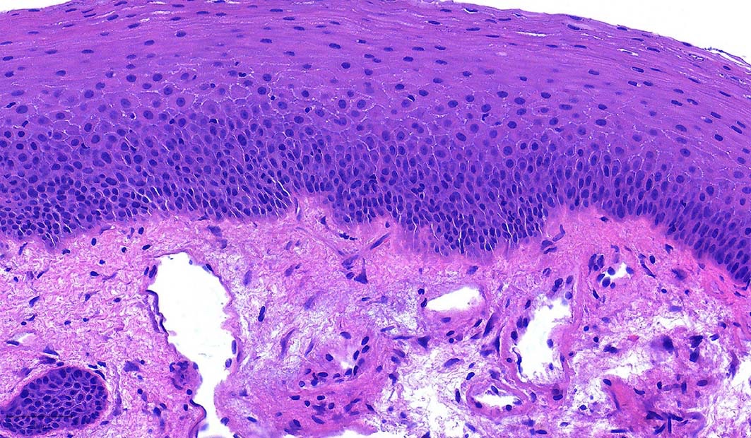
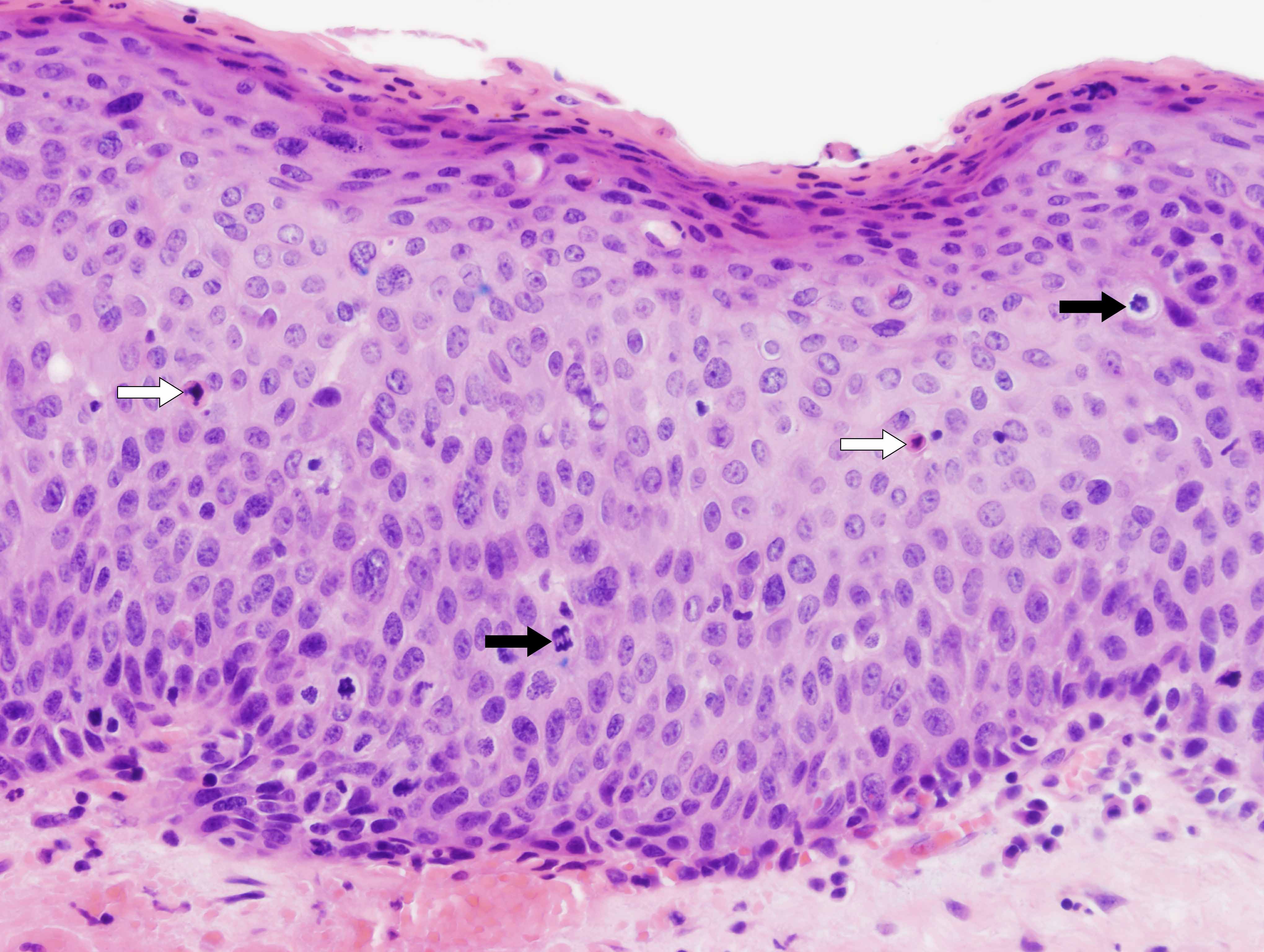
Pathology Outlines Dysplasia

Unilateral vocal cord paralysis a review of CT findings, mediastinal causes, and the course of
Vocal Cord Paralysis Causes, Symptoms, Recovery, Surgery, Treatment
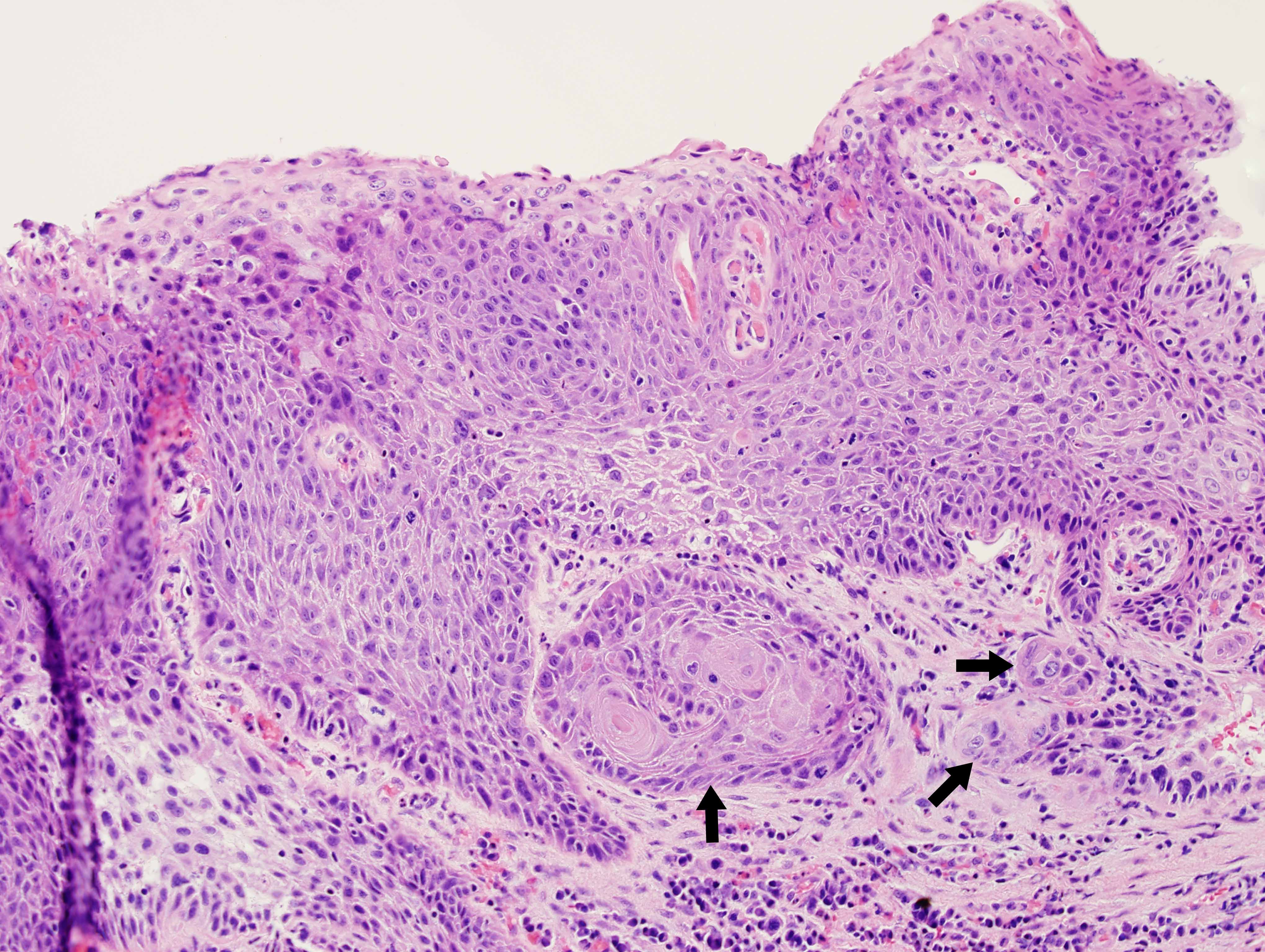
Laryngeal Papilloma with High Grade Squamous Dysplasia Flickr
Vocal Cord Nodules
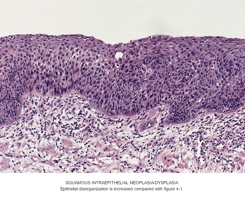
Pathology Outlines Squamous dysplasia

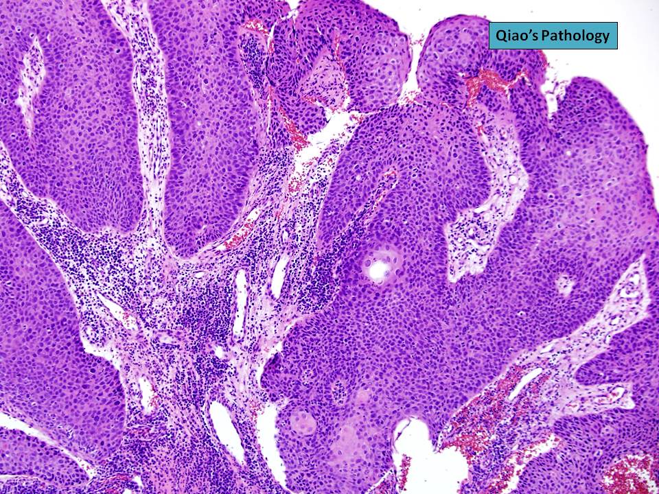
Flickriver Photoset ‘Vocal Cord Polyp’ by Qiao’s Pathology (Art and Science in Medicine)
Pathology Outlines Vocal cord polyp
Invasive squamous cell carcinoma initially presenting as laryngeal leukoplakia Iowa Head and
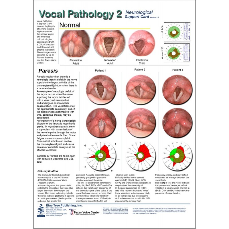
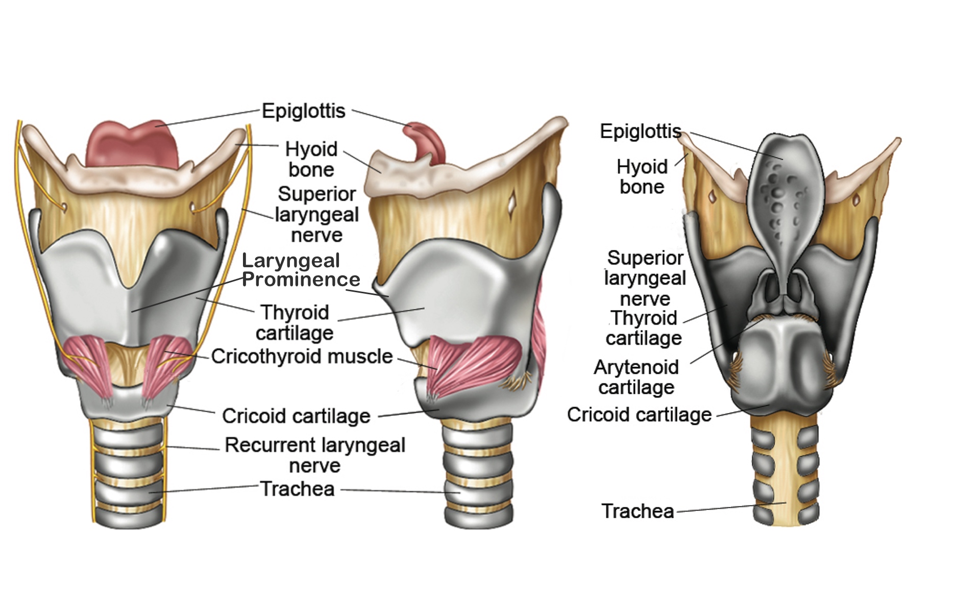
Vocal Pathology II Anatomical Chart
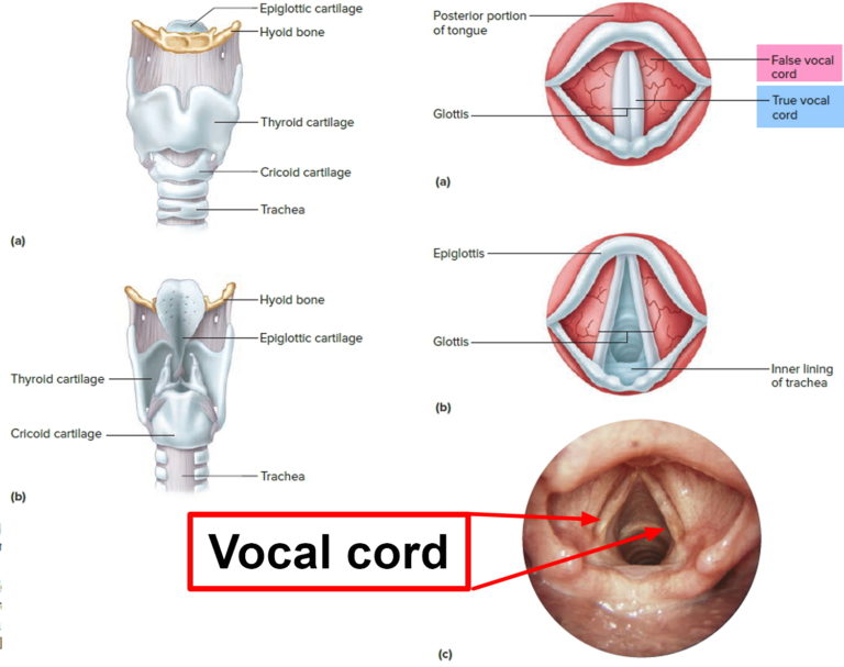
The larynx home of the voice!
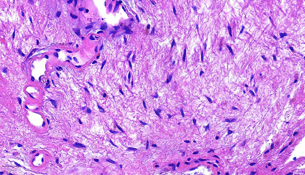
Summary. Cysts of the true vocal cords are less common than other laryngeal cysts. They are usually easily recognized and managed. Patients present with complaints of hoarseness and/or dyspnea. We report our experience with 41 cases of cysts located in the true vocal cords. Clinical and histological aspects are reviewed and discussed.. Vocal-fold leukoplakia and dysplasia are together designated “epithelial hyperplastic laryngeal lesions” (EHLL). Work-up and follow-up are founded on optical examination with high-definition imaging, stroboscopy and narrow-band imaging. Diagnosis is based on pathology, using the new 2017 WHO classification, dichotomizing “low grade” and “high.


